Home

smūgis Puikus ąžuolas Savininkas t1 t2 spine karūna draudimas Minimalus

Preoperative MRI of thoracic spine: T2 (A) and T1-weigh | Open-i

xmlinkhub
xmlinkhub

Intelligent Chiropractic - T1, T2, & T3: The Top Three Thoracic Vertebrae  Tension, Tightness, & Tech Neck Posture are also 3 T's associated with  symptoms that affect your upper thoracic spine. When
Intelligent Chiropractic - T1, T2, & T3: The Top Three Thoracic Vertebrae Tension, Tightness, & Tech Neck Posture are also 3 T's associated with symptoms that affect your upper thoracic spine. When

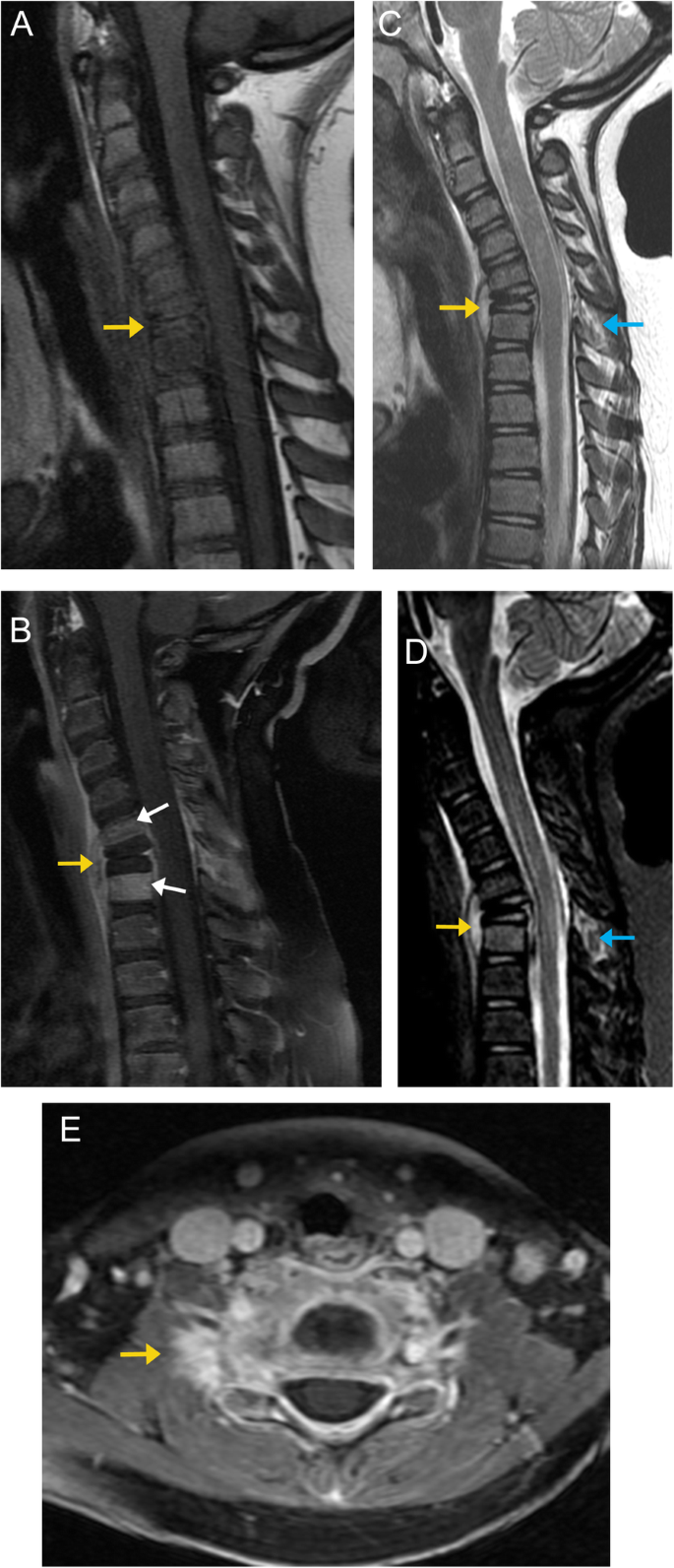
Bone up on spinal osseous lesions: a case review series | Insights into  Imaging | Full Text
Bone up on spinal osseous lesions: a case review series | Insights into Imaging | Full Text

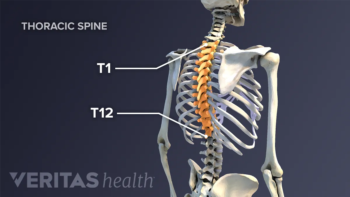
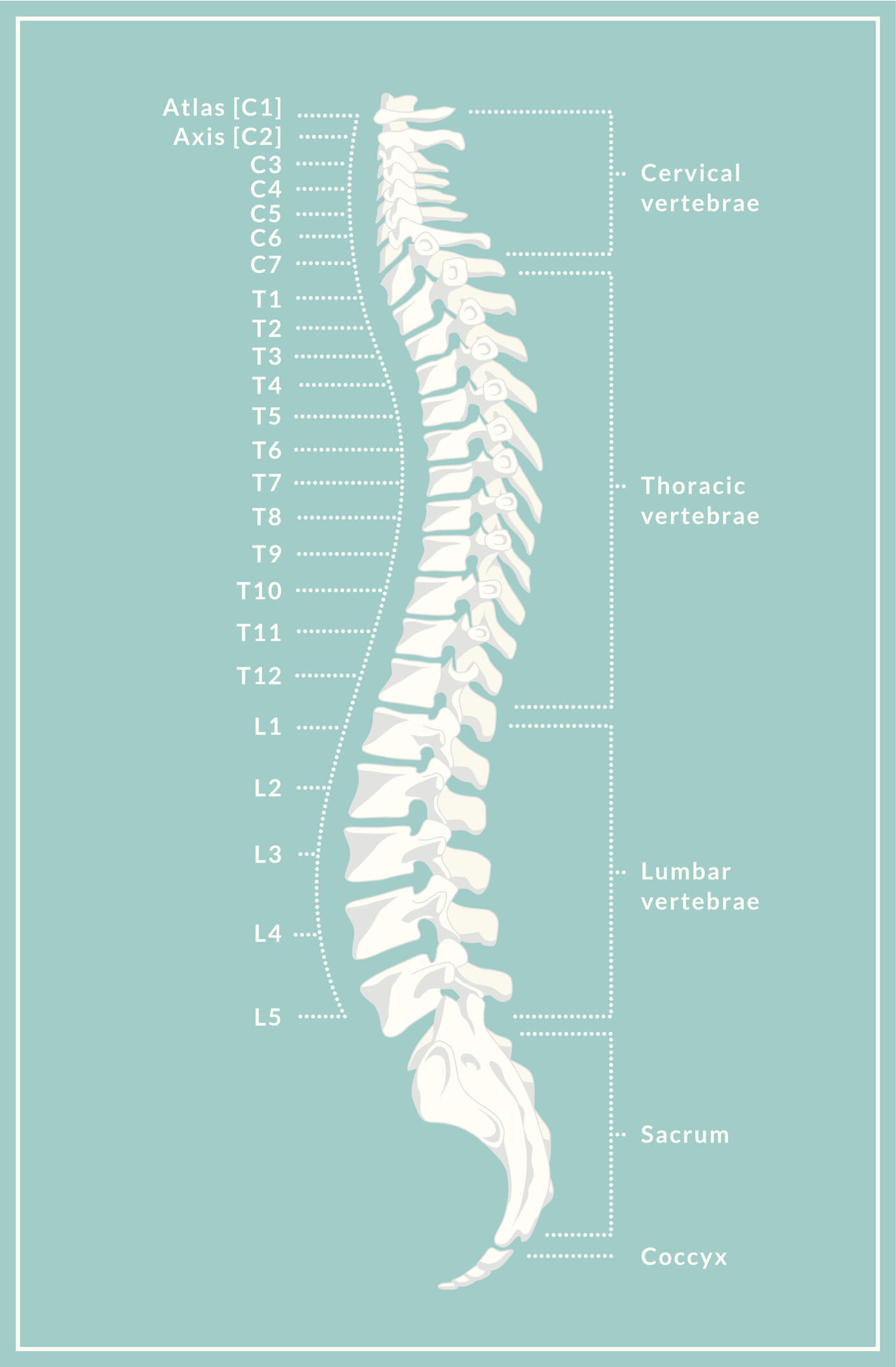
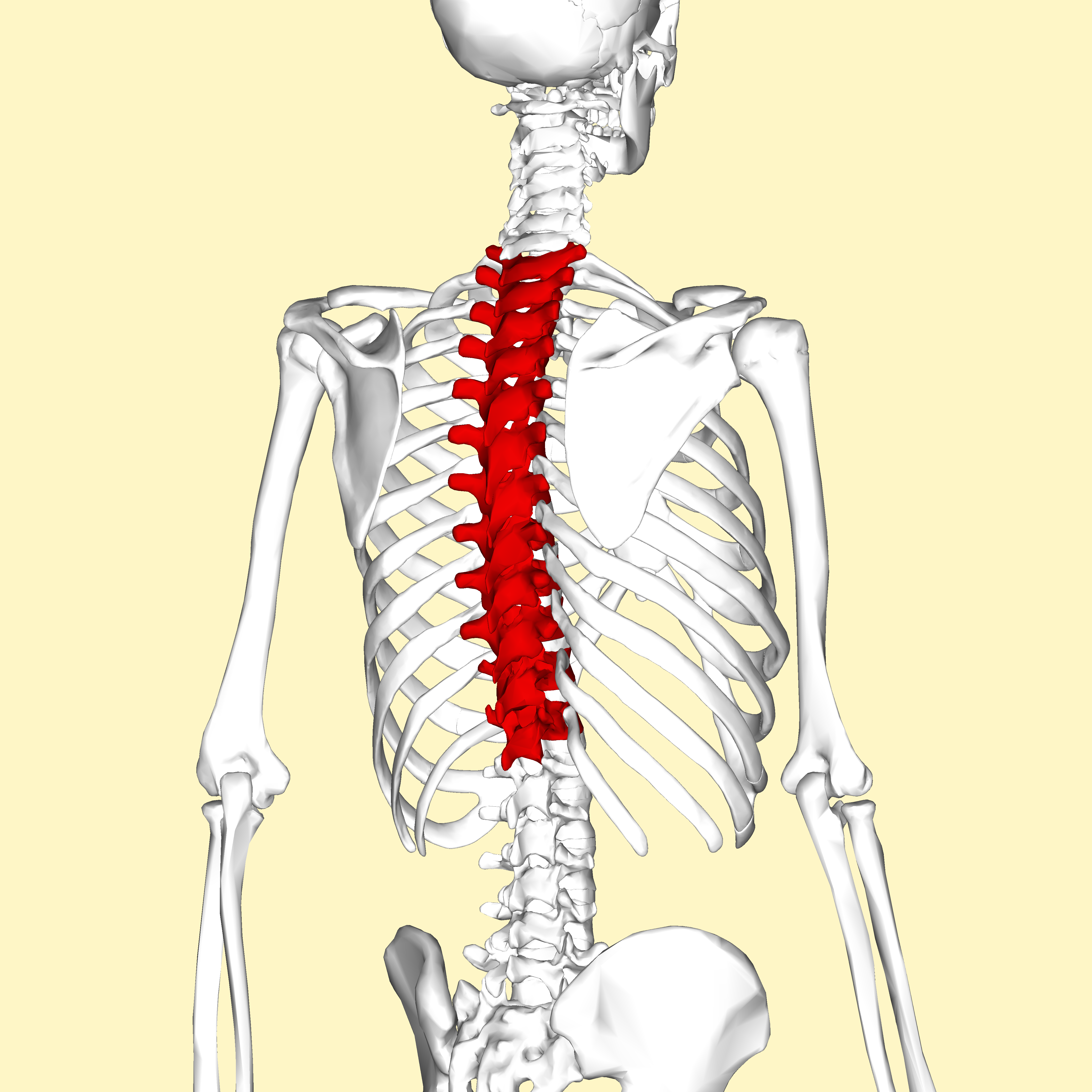
Thoracic vertebrae: Anatomy, function and definition | Kenhub

Comparison of Sagittal FSE T2, STIR, and T1-Weighted Phase-Sensitive  Inversion Recovery in the Detection of Spinal Cord Lesions in MS at 3T |  American Journal of Neuroradiology
Comparison of Sagittal FSE T2, STIR, and T1-Weighted Phase-Sensitive Inversion Recovery in the Detection of Spinal Cord Lesions in MS at 3T | American Journal of Neuroradiology

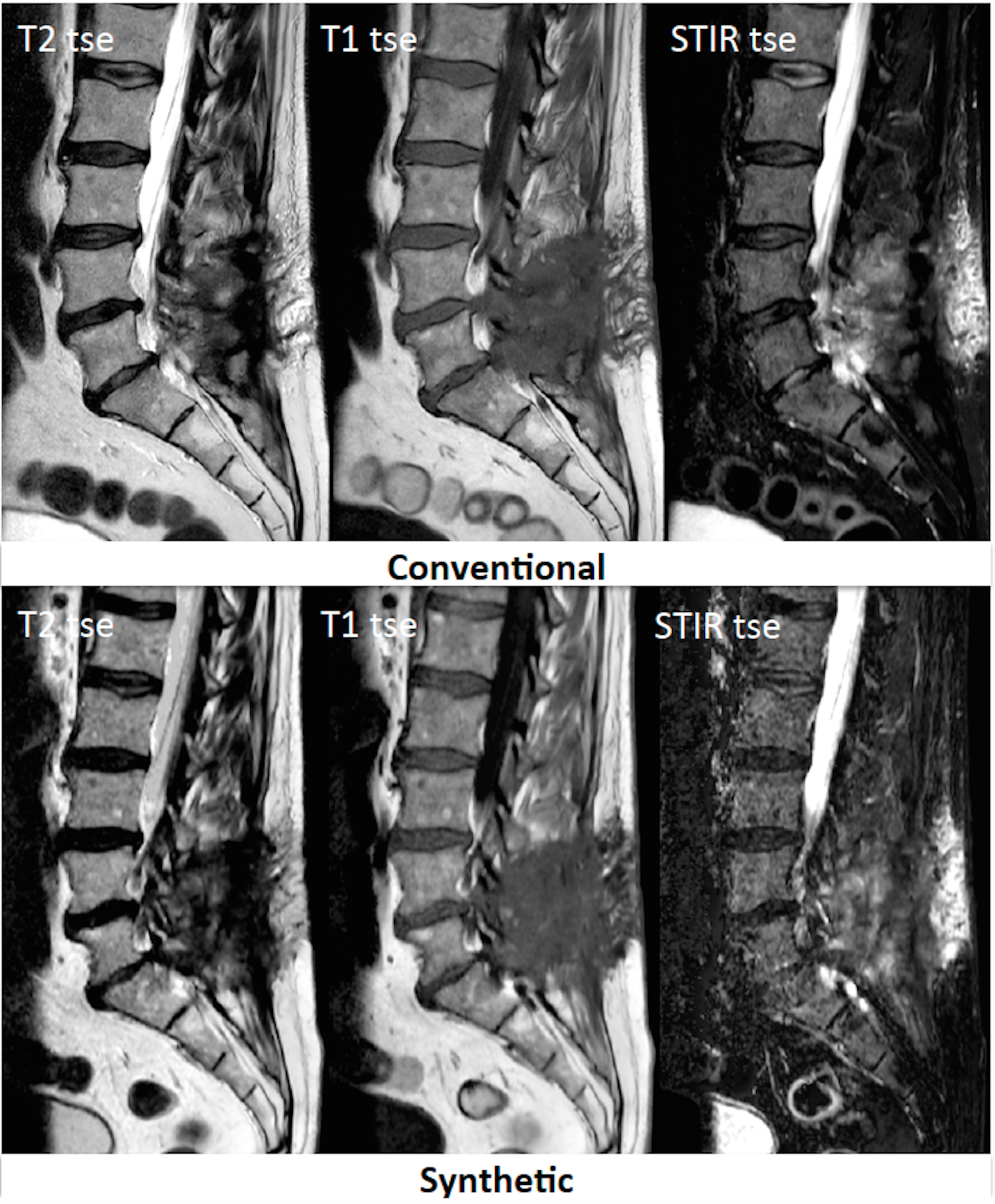
Feasibility of Synthetic MRI of the spine – fast and quantitative imaging

MRI Basics
MRI Basics

Thoracic Spinal Nerves | Spine-health

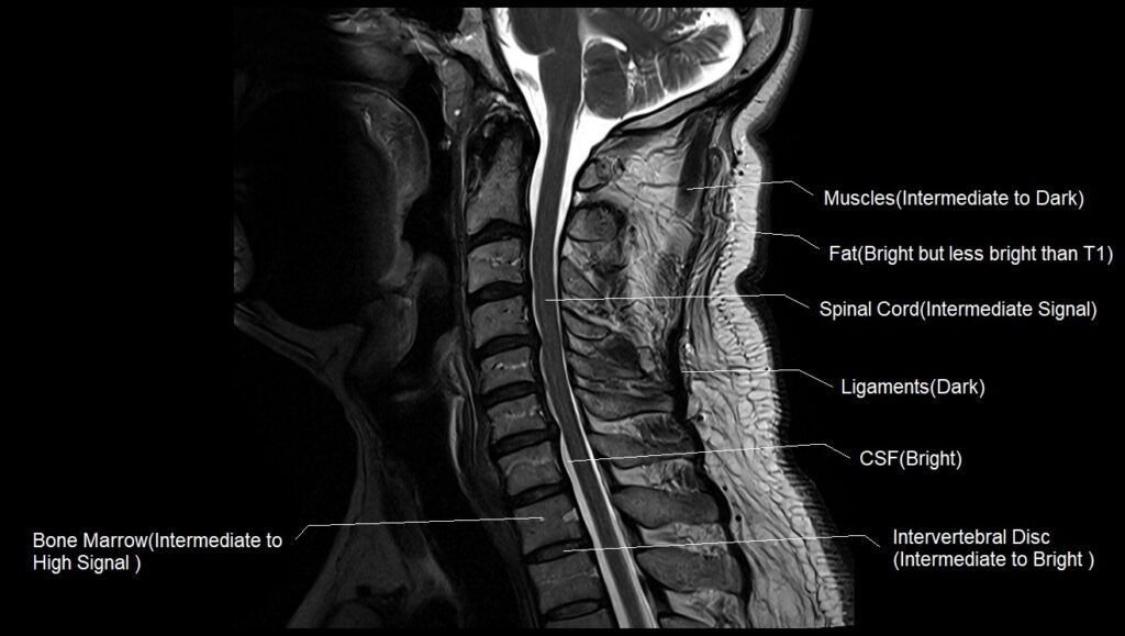
T1 vs T2 MRI | T1and T2 MRI image comparison

Spinal Anatomy Overview - Atlanta Brain and Spine Care

Comparing T1-weighted and T2-weighted three-point Dixon technique with  conventional T1-weighted fat-saturation and short-tau inversion recovery  (STIR) techniques for the study of the lumbar spine in a short-bore MRI  machine - ScienceDirect
Comparing T1-weighted and T2-weighted three-point Dixon technique with conventional T1-weighted fat-saturation and short-tau inversion recovery (STIR) techniques for the study of the lumbar spine in a short-bore MRI machine - ScienceDirect

Thoracic spondylotic myelopathy presumably caused by diffuse idiopathic  skeletal hyperostosis in a patient who underwent decompression and  percutaneous pedicle screw fixation - Shota Miyoshi, Tadao Morino, Haruhiko  Takeda, Hiroshi Nakata, Masayuki Hino,
Thoracic spondylotic myelopathy presumably caused by diffuse idiopathic skeletal hyperostosis in a patient who underwent decompression and percutaneous pedicle screw fixation - Shota Miyoshi, Tadao Morino, Haruhiko Takeda, Hiroshi Nakata, Masayuki Hino,
thoracic spine | Musculoskeletal Key

Pre-contrast T1-weighted imaging of the spinal cord may be unnecessary in  patients with multiple sclerosis | European Radiology
Pre-contrast T1-weighted imaging of the spinal cord may be unnecessary in patients with multiple sclerosis | European Radiology

T1 vs T2 MRI | T1and T2 MRI image comparison

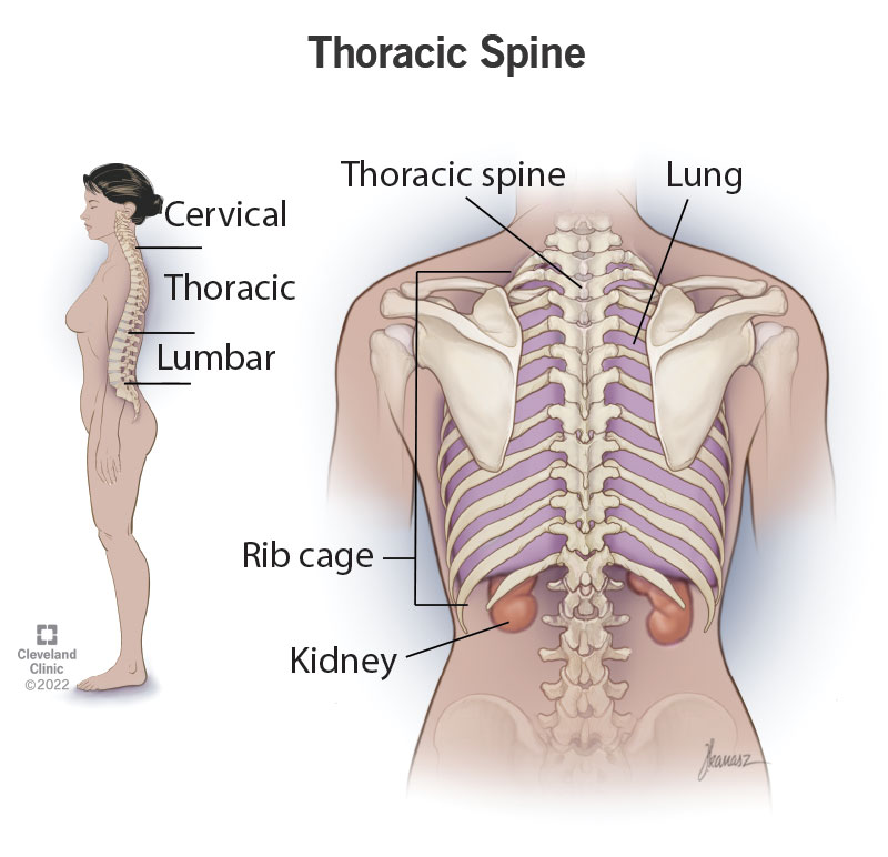
Thoracic Spine: What It Is, Function & Anatomy

Thoracic vertebrae - Wikipedia

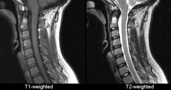
Sagittal T1 and T2 weighted images through the cervical spine... | Download  Scientific Diagram
Sagittal T1 and T2 weighted images through the cervical spine... | Download Scientific Diagram

A Rare Case of T1-2 Thoracic Disc Herniation Mimicking Cervical  Radiculopathy | International Journal of Spine Surgery
A Rare Case of T1-2 Thoracic Disc Herniation Mimicking Cervical Radiculopathy | International Journal of Spine Surgery
Thoracic Spine Fractures & Injuries | Shepherd Center

MRI interpretation - T1 v T2 images

What do T1 and T2 weighted images mean in an MRI? - Quora

Anatomy of the lumbar vertebrae: normal anatomy | e-Anatomy

Comparison of MRI T1, T2, and T2* mapping with histology for assessment of  intervertebral disc degeneration in an ovine model | Scientific Reports
Comparison of MRI T1, T2, and T2* mapping with histology for assessment of intervertebral disc degeneration in an ovine model | Scientific Reports